Dr. Kapadia regularly treats patients with cancers involving the eyelids. Eyelid reconstruction is not cosmetic surgery and not everyone has a perfect result. Nonetheless, most patients are very pleased with their outcome and show minimal evidence of surgery after they heal. Approximately 10% of all skin cancers occur on the eyelids. As in other parts of the body, basal cell carcinoma is the most common type of eyelid skin cancer.
Patient 1 - Basal cell carcinoma of the right upper eyelid, before and after
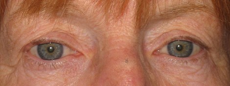
A. Before surgery
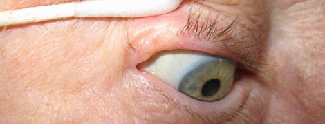
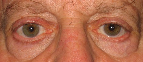
B. Before surgery, magnified view
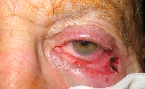
C. After excision of tumor
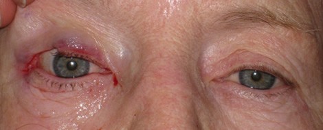
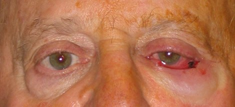
D. Immediately after surgery
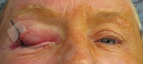
E. Six weeks after surgery
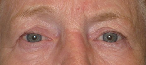
These photos show a woman in her 70s with a basal cell carcinoma of the right upper eyelid. Figure A shows relatively subtle changes in the lateral aspect of the right upper eyelid. The magnified view in Figure B demonstrates multiple fine blood vessels (telangectasis) and loss of eyelashes. A biopsy of this area was positive for basal cell carcinoma. The patient underwent Mohs micrographic surgery to remove the tumor and was left with a large defect of the lateral right upper eyelid, just under 1/2 of the total eyelid width (Figure C). Figure D shows the appearance of the eyelid immediately after reconstructive surgery. After several weeks, the eyelid is well healed and there is minimal visible signs of the prior cancer or reconstructive surgery. Careful examination of the photo shows a subtle change in the contour of the right upper eyelid compared to the other side which should improve after several months of healing.
Patient 2 - Basal cell carcinoma of the left lower eyelid
A. Before Surgery
B. After Excision of Tumor
C. After excision of tumor, magnified view
D. Five months after surgery
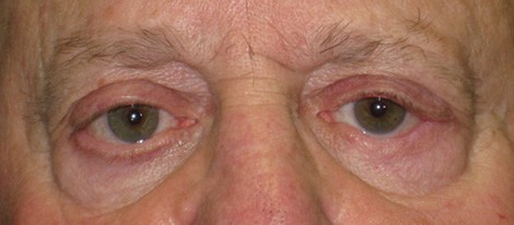
These photos show a man in his 70s with recurrent basal cell carcinoma of his left lower eyelid. He underwent excision of a basal cell carcinoma and reconstructive surgery on that eyelid five years prior and noticed increasing "lumpiness" of that eyelid. Figure A shows irregularity of the lateral portion of the left lower eyelid. An eyelid biopsy was performed, confirming the diagnosis of recurrent basal cell carcinoma. Figures B and C show the appearance of the eyelid after tumor resection. The defect encompasses approximately 2/3 of the normal eyelid width. Extensive reconstruction was performed, which included borrowing tissue from the upper lid and obtaining a skin graft from the shoulder area. Five months after surgery, the eyelid is well healed and shows only a subtle difference compared to the other side.