Basal cell carcinoma is the most common form of eyelid skin cancer, but there are many other types of eyelid cancers including squamous cell carcinoma, sebaceous cell carcinoma and melanoma. Not all patients have a perfect result after this type of surgery, but most patients are very pleased with their overall result.
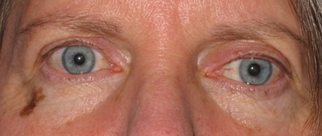
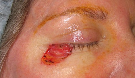
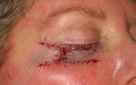
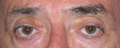
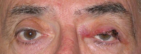
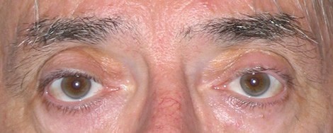
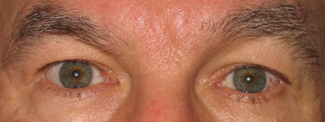
Patient 1 - Melanoma of the right lower eyelid and right cheek
A. Before surgery
B. After excision of tumor
C. After eyelid reconstruction
D. Six months after surgery
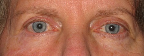
This patient had a pigmented area below her right eye which had been stable in size and appearance for more than 30 years. A biopsy was performed after the lesion starting increasing in size and showed lentigo maligna, a superficial form of malignant melanoma. The lesion was excised by Mohs surgery and reconstruction was performed using advancement flaps to bring adjacent skin into the defect. At six months, the eyelid is well healed with only minimal evidence of surgery. Careful examination shows that the right lower eyelid is in a slightly lower position than the eyelid on the left side.
Patient 2 - Squamous cell carcinoma of the left lower eyelid
A. Before Surgery
B. After Excision of Tumor
C. Three months after surgery
This patient had a small area of redness and scaling of the left upper eyelid which was barely visible in the preoperative photo. A biopsy of this area was positive for squamous cell carcinoma. The tumor was excised by Mohs surgery and reconstruction was performed with a skin flap to bring adjacent tissue into the defect. The eyelid is fully healed three months after surgery.
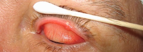
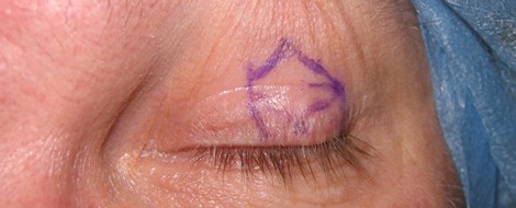
Patient 3 - Sebaceous carcinoma of the eyelid
A.Before surgery
B. Before surgery, magnified view
C. Before surgery, eyelid marked for excision
D. Three months after surgery
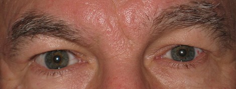
These photos show a patient with sebaceous cell carcinoma, a rare but serious form of eyelid cancer. The lesion appeared as a mass on the margin of the eyelid similar to a chalazion or stye. Biopsy of the lesion showed sebaceous cell carcinoma of the eyelid. A large portion of the eyelid was excised in entirety and the edges of the eyelid brought back together. Three months after surgery, the eyelid has a normal appearance. The edge of the wound is covered by excess skin, but the eyelid appears normal even when this excess skin is lifted away (photo not shown). Because sebaceous cell carcinoma may metastasize to other parts of the body, this patient also had a lymph node biopsy which did not find any evidence of tumor spread.